Chest Wall Tumour: Causes, Symptoms, Signs, and Treatment

Medically Reviewed By
Dr. Ragiinii Sharma
Written By Srujana Mohanty
on Oct 31, 2022
Last Edit Made By Srujana Mohanty
on Mar 18, 2024

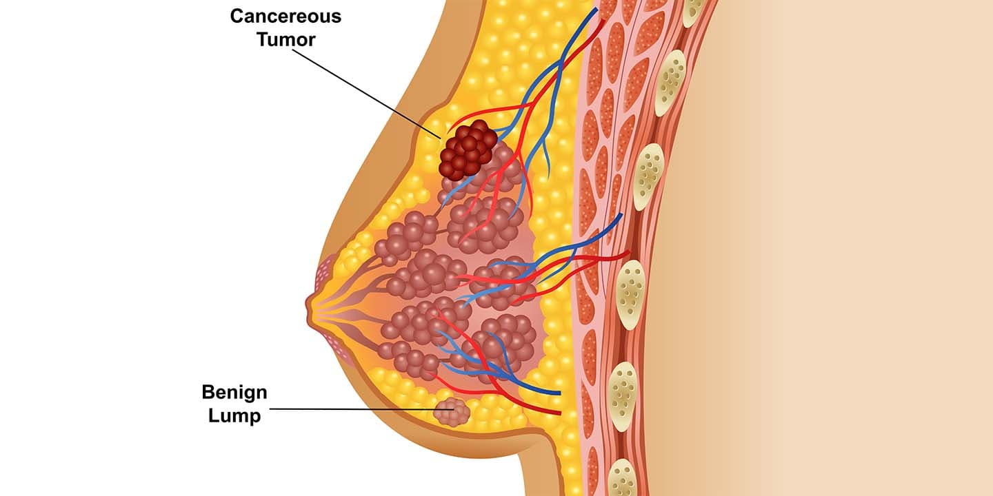
Tumours are the cellular masses formed by the multifold division of an abnormal group of cells manifesting into cancerous or non-cancerous lumps. The tumours developed in the protective wall of the heart, lungs, and liver are commonly referred to as “chest wall tumours.”
The chest wall forms the protective wall against the soft tissues and organs present in the chest, including the heart, lungs, and liver. The chest wall is primarily composed of soft tissues, bone, and cartilage.
The cancerous and non-cancerous tumours can grow in the chest wall and may or may not show signs. Moreover, these tumours are of different types, and their symptoms also vary making it difficult to list all symptoms.
This article explores everything you need to know about chest wall tumours, their types, symptoms, diagnosis, and treatment.
What is a chest wall tumour?
Tumours are defined as the abnormal growth of cells that can be either malignant (cancerous) or benign (non-cancerous). Like any other cancer, the tumours developed in the chest wall are commonly called “chest wall tumours.”
The tumours can develop either in the cartilage, bone, or soft tissues of the chest wall. Sometimes, the tumours developed in these regions can interfere with pulmonary function and also metastasise (spread) to the internal tissues/ organs of the chest region.
The most common forms of chest wall tumours are seen in the bones or ribs and the cartilage. It is estimated that nearly 60% of the chest wall tumours that develop manifest into cancerous growths.
The tumours in the chest wall can be either of two types,
- Primary chest wall tumours: The primary chest wall tumours originate in the muscles or bones of the chest wall. Most of the primary chest wall tumours are non-cancerous. And about 5% (or 1 in 50) of all tumours in the chest are primary chest wall tumours.
- Secondary chest wall tumours: The tumours that have developed elsewhere in the body and spread to the chest wall. Secondary chest wall tumours are always malignant. The secondary chest wall tumours spread from other organs, including the breasts, colon, lungs, prostate glands, stomach, thyroid, uterus, and kidneys.
The cancerous growths of the chest wall can get aggressive, spreading through the internal organs of the lungs and liver. And hence, keeping a track of possible symptoms and early diagnosis can help prevent tumour advancement while promoting a good prognosis with early treatment.
Types of chest wall tumour
The primary non-cancerous chest wall tumours can be classified into the following types depending on their occurrence and tissue of origin.
- Cavernous hemangioma: It is a type of soft tissue tumour that is mostly seen in children.
- Chondroma: It is the type of tumour that occurs in the cartilage.
- Desmoid tumour: It is the form of recurring tumour seen in the soft tissue.
- Eosinophilic granuloma: It affects the bone and usually occurs in the shoulder blade or collarbone.
- Fibrous dysplasia: It is the type of tumour that forms in the ribs and a common non-cancerous type of chest wall tumour.
- Chest wall lipoma: It is a form of non-cancerous tumours forming in the soft tissues.
- Lymphangioma: It originates in the small tissues and is commonly seen in children.
- Myxochondroma: A form of primary benign tumour that develops in the cartilage.
- Osteochondroma: it is one of the most common types of primary non-malignant tumour formed in the cartilage.
The primary cancerous chest wall tumours are categorised into the following types.
- Chondrosarcoma: This malignant form of chest wall sarcoma is formed in the cartilage and is usually one of the aggressive and most common forms that can metastasise (spread) to the ribs or breastbone (sternum).
- Ewing’s sarcoma: It usually forms in bone or soft tissue surrounding the bone. Typically seen in young adults and children and majorly affects one single bone or rib and is the most commonly occurring chest wall tumour type.
- Fibrosarcoma: It is another form of soft tissue malignant chest wall tumour type, primarily affecting young adults.
- Liposarcoma: As the name goes, this form of cancerous tumours originates in the fat cells or tissues.
- Malignant fibrous histiocytoma: Majorly affecting older adults in their 50s and 60s, this primary cancerous chest wall tumour is formed in the soft tissues.
- Myeloma: This type of chest wall tumour originates in the bone of adults above 40.
- Osteosarcoma: It is the most aggressive form of chest wall cancer that forms in the bone.
- Rhabdomyosarcoma: Though commonly seen in children and adolescents, it typically forms in the soft tissues.
Chest wall tumour signs and symptoms to look out for
Chest wall tumours can affect anyone at any age. Some types are common in children, while some are typical among older adults.
Chest wall tumours affecting younger adults or children are likely to be small and non-cancerous forms. However, in people above 40, the chest wall tumours are likely to be large and malignant.
Chest wall tumour symptoms may be difficult to identify as they can be relative to symptoms of other medical conditions. The symptoms vary depending on the type of tumour. Some do not even realise these symptoms until the tumours have outgrown their advanced stage.
Here are some noticeable signs and symptoms of chest wall tumours you need to be wary of.
- A protruding mass on the chest wall,
- Visible bumps or lumps on the chest wall,
- Swelling, tenderness, and soreness in localised regions of the chest,
- Chronic pain in the chest,
- Muscle atrophy (breakdown),
- Unexplained loss of weight,
- Pain while moving the body,
- Impaired movement,
- Difficulty in breathing.
What causes chest wall tumours?
There is no clear-cut reasoning as to what causes chest tumours. However, scientists believe that,
- Genetics,
- Diet, and
- Lifestyle factors play a dominant role in the transposition of these tumours, particularly secondary chest wall tumours.
Tests to diagnose chest wall tumours
Diagnosis of chest wall tumours involves a thorough review of medical history and symptoms. The initial tests include imaging tests, and biopsies that your healthcare provider will order if you have symptoms, signalling a chest wall tumour.
Some imaging tests that help detect chest wall tumours include:
- Chest X-ray: The chest x-ray provides a prompt evaluation of the lung for any direct invasion. It may detect cortical bone destruction or confirm whether the tumour originates from the bone or not.
- Computed tomography (CT) scan: A more sensitive test than the chest x-ray and aids verification of x-ray diagnosis.
- Magnetic resonance imaging (MRI): MRI helps evaluate the soft tissues and determines the chest wall structural abnormalities of infection, lesions, or inflammation.
- Positron emission tomography (PET) scan: PET evaluates the chest wall tumour stage and its response to treatment while detecting the disease recurrence possibilities.
Though imaging tests can detect the presence of tumours, a needle or an open biopsy can help confirm a definitive diagnosis and determine if these growing tumours are malignant or benign.
Chest wall tumour treatment
Treatment for chest wall tumours depends on the type of tumour developed, the location of the tumour, and the associated medical conditions of the patient.
Here is a basic outline of treatment options considered for chest wall tumours.
- Surgery: Surgery can be considered in cases of benign and malignant chest wall tumours.
Benign tumours can be surgically removed to ease movement, allow proper organ functioning, and reduce muscle wearing out. And in cases where the tumour is malignant, treatment involves a combination of surgical removal, reconstruction with chemotherapy, and radiation therapy.
During surgical removal and reconstruction, the affected portion of the rib cage is removed and replaced with mesh or “fibre” material. This allows effective repair of the tumour site.
- Radiation therapy: This therapy after surgical reconstruction of malignant tumours uses high-intense radiation to kill cancer cells and slow down their growth.
- Chemotherapy: Chemotherapy for chest wall tumours uses drugs that nullify the growth of cancer cells. These chemotherapeutic drugs can be given orally, intravenously, by infusion, or through the dermis based on the cancer stage and type. Sometimes, chemotherapy may be recommended before surgery, which helps reduce the tumour size before its removal.
Frequently Asked Questions
- What type of chest wall tumours is more common?
Non-cancerous chest wall tumours are more common than cancerous chest wall tumours. They are detected and treated when they interfere with other organs functioning, causing breathing problems or pain. Non-cancerous tumours that are more common include osteochondroma and fibrous dysplasia.
- What is the survival rate of chest wall cancer?
The prognosis of chest wall cancer varies depending on the cancer type, cell differentiation, and staging. Sarcomas are the most aggressive and studied type of chest wall tumour. According to statistical reports, around 17% of primary chest wall sarcomas are reported to have a 5-year survival rate. However, survival rates can increase if the disease is diagnosed early.
- Is cancer in the chest curable?
Non-cancerous chest wall cancers are highly treatable. Cancerous chest wall tumours are also treatable when screened early. Around 80 to 90% of early-stage or small primary or secondary chest wall cancers are curable. However, these rates drop with cancer advancement and metastasis to other body parts.
Conclusion
Chest wall tumours refer to the tumours developed in the chest wall or the protective barrier of the chest. They can be both cancerous and non-cancerous. The prognosis and treatment of the cancerous type widely depend on the tumour type and how far it has spread.
Early detection of these cancerous tumours can give a healthy long-term outlook and optimise the likelihood of successful treatment and good quality of life. The chest wall cancer treatment ranges from reconstruction and cancer removal surgeries to chemo and radiation therapy.
It is always advisable to get connected with a doctor if you have one or more chest wall tumour symptoms for an early screening and treatment procedure.


